Development of Mice Model for the West Nile Virus (Strain Mor/1996)

Abstract
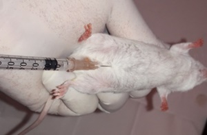
The present study aims to develop and validate a mouse model for in vivo infection with West Nile Virus (WNV). To achieve this, 90 non-vaccinated native albino mice were divided into three groups according to three modes of inoculation: Intracerebral (IC), Subcutaneous (SC), and Intraperitoneal (IP). Each group consisted of 25 challenged animals and five controls. After inoculation, daily observation of clinical symptoms was conducted based on a predetermined scoring chart over a period of 2 months. After the animals' deaths, autopsy and decerebration were performed for histopathological analysis and an RT-PCR test. The results showed that the most comprehensive clinical presentation was observed in animals inoculated via the IP and SC routes, including ataxia, weight loss, dehydration, signs of constipation, fasciculation and blindness. However, for the IC route, the vast majority of mice suddenly died without exhibiting clinical signs, apart from ataxia. The mortality rate among inoculated animals was 96% for the IC route, 56% for the IP route and 44% for the SC route. Histopathological examination revealed non-pathognomonic but well-marked signs such as discrete to moderate focal perivascular gliosis and mild to moderate neuronal degeneration and necrosis. On the other hand, all samples tested by RT-PCR were positive for WNV. Furthermore, the calculation of the LD50 revealed values of 104.3 and 104.8 for IC and IP routes respectively. It results from this study that the validated inoculation routes for the mouse model of West Nile virus infection are the IP and SC routes.
Keywords: West Nile, Mice- Model, Challenge, RT-PCR